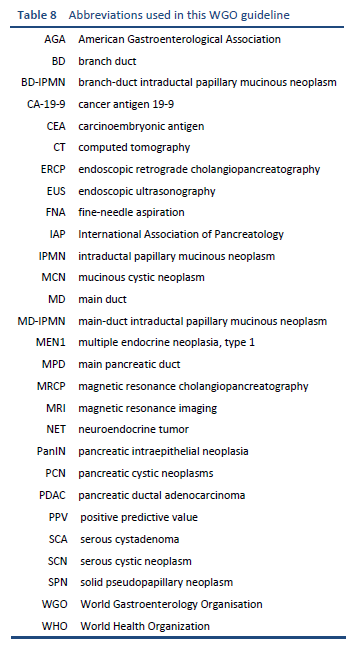
World Gastroenterology Organisation

Global Guardian of Digestive Health. Serving the World.


WGO Practice Guideline:

Pancreatic Cystic Lesions


March 2019


Review Team

Juan Malagelada, Chair (Spain)
Nalini Guda, Co-chair (USA)

Khean-Lee Goh (Malaysia)
Thilo Hackert (Germany)
Peter Layer (Germany)
Xavier Molero (Spain)
Stephen Pandol (USA)
Masao Tanaka (Japan)
Muhammed Umar (Pakistan)
Anton LeMair (Netherlands)


Contents

(Click to expand section)

1.  Cascades and Key Points In Diagnosis And Management

1.1  Key Points and Main Practice Statements

  • A careful patient history and physical evaluation are necessary.
  • At the initial cyst fluid aspiration: carry out carcinoembryonic antigen (CEA), amylase, and cytology testing.
  • Cysts should be assessed to determine the risk for malignancy or complications.           
    In patients with symptomatic cysts, cysts larger than 3 cm, with solid components, or with ductal dilation:
    — Perform endoscopic ultrasonography (EUS) with or without fine-needle aspiration (FNA)* and consider surgical evaluation.
    In patients who have cysts with low-risk features:
    — Continue noninvasive surveillance for at least 5 years, on the basis of some of the guidelines, although the longer-term risk for the development of malignancy is not very clear.    
    — Consider the patient’s age and comorbidities and continue surveillance as appropriate until there is better evidence for definite discontinuation of surveillance and risk stratification.
    — The patient’s preferences and understanding of the risk should be taken into consideration.
    In patients who have indeterminate cysts:
    — Surveillance with cyst fluid analysis and/or imaging features.
  • Molecular testing is not routinely done because of limited data and the expense, but it does hold promise for the future.
  • A thorough discussion with the patient regarding the diagnosis, current dilemmas in diagnosis and treatment, and the economic and emotional burden of investigations should be conducted before initiating any surveillance strategy.

* FNA may not be performed if the lesion is in the pancreatic body or tail if there is any concern about malignancy, due to the risk of seeding along the FNA track that may not be addressed by subsequent surgery. In patients who have lesions in the body or tail, the stomach is not removed, whereas in those with lesions in the pancreatic head, the duodenum is removed at surgery.

1.2  WGO Cascades For Pancreatic Cystic Lesions

WGO cascades: a hierarchical set of diagnostic, therapeutic, and management options to deal with risk and disease, ranked by the resources available.

WGO guidelines and cascades are intended to highlight appropriate, context-sensitive and resource-sensitive management options for all geographical areas, regardless of whether they are “developing,” “semi-developed,” or “developed.” WGO cascades provide options that are not necessarily defined solely by resource priorities and may, for example, also include cost–benefit factors, patient preferences, and the availability of equipment, skills, and expertise.

Most asymptomatic incidental cysts are diagnosed in resource-enabled countries when imaging is being carried out to evaluate symptoms not necessarily related to pancreatic disease. For the Asian–Pacific region, for instance, two recent papers from Korea and Japan—both affluent countries—report asymptomatic cyst incidence rates of 2.2% and 3.5% [1,2]. In low-resource countries, most diagnoses are established at surgery or autopsy.

The authors of this guideline have therefore chosen not to use the conventional “cascade” pattern, but rather to make recommendations based on the current evidence. We understand that all the resources are not available everywhere, and an informed decision should be made in discussion with the patient regarding the risk for malignancy, available resources, and cost.

2.  Introduction

2.1  Scope and Goals

This guideline aims to provide physicians worldwide with a reasonable, up-to-date approach to the management of pancreatic cystic lesions. Since pertinent diagnostic and therapeutic resources are not uniformly available in different areas of the world, these guidelines are meant to be used as appropriate, with local resources and patient preferences being kept in mind.

2.2  Definitions

“Pancreatic cystic lesions” is a conventional term that refers to a well-defined lesion in the pancreas that contains fluid. Most small lesions are detected incidentally when scanning is performed for evaluation of non–pancreas-related indications or symptoms. The etiology of pancreatic cysts is variable; they may be inflammatory or posttraumatic, or may have no known etiology. While most small lesions are benign, some lesions can lead to malignancy and hence a need for further work-up, surveillance, and management decisions. It is therefore necessary to obtain a good patient history and assess the nature of the lesion through appropriate investigations as needed, in order to assess the risk for malignant progression. Since potentially malignant lesions cannot be distinguished reliably from benign lesions on the basis of the clinical and morphological features alone, further evaluation and/or surveillance may be necessary.

Premalignant cystic lesions of the pancreas include mucinous cystic neoplasms and intraductal papillary mucinous neoplasms. As indicated above, some pancreatic cystic lesions may evolve into adenocarcinoma of the pancreas [3].

Since pancreatic ductal adenocarcinoma (PDAC) and pseudopapillary tumors rarely present as cystic lesions, they are not covered in the present guideline.

Pancreatic pseudocysts, which lack a definite cyst wall, usually occur in patients who have a history of pancreatitis or trauma. Pseudocysts are benign and often resolve spontaneously without any need for intervention, unless they are symptomatic; they are not the subject of the current guideline. However, it is important to ensure that the lesion is in fact a pseudocyst and not a true pancreatic cyst. While the management of benign or obviously malignant lesions is less ambiguous, treatment for indeterminate-risk or intermediate-risk lesions is unclear, and these guidelines will hopefully provide guidance on the appropriate work-up and management.

3.  Cystic Lesions of the Pancreas

Pancreatic cysts are often asymptomatic; they are often benign, but some have malignant potential.

Pancreatic cystic lesions may be classified as:

  • Benign cysts—e.g., simple cysts, pseudocysts, and serous cystic neoplasms (SCNs)
  • Cysts with malignant potential—e.g., pancreatic cystic neoplasms (PCNs) such as mucinous cystic neoplasms (MCNs) and intraductal papillary mucinous neoplasms (IPMNs)
  • Malignant cysts: neoplastic cysts such as pancreatic adenocarcinomas with cystic degeneration, and cystic pancreatic neuroendocrine tumors

Pancreatic cystic neoplasms may have a malignant potential; these include MCNs and IPMNs. Alternatively, they may have no malignant potential; these include serous cystic neoplasms (SCNs). Benign cystic lesions may be managed conservatively, while those with a significant malignant potential need surgical intervention [4].

3.1  Classification of Pancreatic Cystic Lesions

Table 2 presents the World Health Organization (WHO) histological classification of pancreatic cysts, which also includes solid pseudopapillary neoplasms.

Origin of lesions—relevant to management of precursor lesions:

  • From progression of an intraductal papillary mucinous neoplasm (IPMN)
  • From progression of a mucinous cystic neoplasm (MCN)

3.2  Pancreatic Intra-Epithelial Neoplasm Panin

Many would agree that the main difference between IPMN and PanIN is size: PanIN lesions are microscopic flat or papillary lesions that arise in the small intralobular pancreatic ducts, usually measure less than 5 mm in size, very rarely form cystic structures, and are commonly undetectable on cross-sectional imaging or EUS. Maire et al. [14] tried to correlate the EUS findings with histopathology in a selected population. When the lesion is larger than 10 mm, IPMN is the preferred term, while below 10 mm the term “dilated side branch” is appropriate. Doubts have been expressed regarding the histological characterization of lesions between 0.5 and 1 cm. Molecular, genetic, or epigenetic markers may be helpful for differentiating between PanINs and IPMNs [3,14].

3.3  Pancreatic Cysts Differential Diagnosis

The following list highlights particularly common potentially confusing appearances that need to be considered in the differential diagnosis:

  • Chronic pancreatitis, versus intraductal papillary mucinous neoplasms
  • Postpancreatitis pseudocysts, versus serous neoplasms, versus mucinous cystic neoplasms
  • Serous cystic neoplasms, versus branch-duct intraductal papillary mucinous neoplasms, versus acinar cell cystadenoma
  • Solid variants of serous cystic neoplasm (SCN), versus neuroendocrine tumor, versus solid pseudopapillary tumors
  • Cystic forms of any solid tumor
  • Rare pancreatic or peripancreatic cystic lesions (e.g., epithelial cysts)


4.  Clinical Presentation

Most pancreatic cysts are asymptomatic and are discovered incidentally on diagnostic imaging that is carried out for an unrelated symptom or reason. In a minority of cases, the initial presentation may be due to a symptomatic cyst manifesting as acute pancreatitis, bleeding, jaundice, or palpable mass. In areas of the world in which advanced diagnostic imaging technology is unavailable or is applied with more restricted criteria, pancreatic cystic lesions may be discovered at a later stage, but this usually implies a larger size or progression to neoplasia.

In patients with a symptomatic cyst, pain is the most common manifestation. Pain may alert the physician to a greater likelihood for malignancy, except in postpancreatitis pseudocysts, and the risk of malignancy may be related to the duration of symptoms [15,16]. Other symptoms include jaundice, nausea, and vomiting secondary to compression of the stomach, or gastric outlet obstruction secondary to extrinsic compression of the duodenal lumen.

Patients with MCNs may also present with pain, an abdominal mass, or weight loss that may have been present for years before the diagnosis [17]. However, most MCNs are discovered on cross-sectional imaging in otherwise asymptomatic patients.

Patient interview: history and background

  • Reasons for consultation
  • Demographic parameters
  • Family and personal history, including pancreatic diseases (e.g., pancreatic cancer, pancreatitis, diabetes)
  • Alcohol use, smoking, drugs, medication
  • Body mass index

Potential clinical presentations related to specific types of cyst: it should be noted that most patients with pancreatic cysts are asymptomatic.


5.  Assessment of Cystic Lesions

5.1  Introduction

The increasing numbers of patients all over the world with incidentally discovered cysts require further refinement of the recommendations for which imaging should be carried out [6,8].

The approach to pancreatic cysts continues to be problematic, due to the lack of good natural history data, few published studies with long-term follow-up data, and possible bias, since most of the reports are from centers that specialize in the treatment of pancreaticobiliary disorders.

  • In general, patients with smaller lesions (< 2 cm) are less likely to be symptomatic, and the lesion is often discovered incidentally.
  • Generally, smaller cystic lesions are less likely to present with concerning radiographic features of malignancy (e.g., solid components or ductal dilation) than larger cysts (> 3 cm).
  • The prevalence of pancreatic cysts increases with age, in part due to increased radiographic surveillance generally performed in older patients with more frequent comorbidities (such as a personal history of malignancy). In most cases, cystic lesions are therefore detected incidentally when computed tomography (CT) and magnetic resonance imaging (MRI) are performed for other reasons.
  • In 95% of the cases, the spectrum of cystic neoplasia includes intraductal papillary mucinous neoplasia (IPMN), mucinous cystic neoplasia (MCN), serous cystic neoplasia, and solid pseudopapillary neoplasia (SPN).
  • The risk of malignancy ranges from 0% to more than 60%. Diagnostic procedures therefore aim to distinguish between neoplastic cystic and nonneoplastic cystic lesions, and also between serous and mucinous lesions, as these have different malignant potentials.
  • A precise diagnosis is required so that the surveillance or therapeutic strategy can be adapted accordingly. This can be provided by an analysis of the imaging data in combination with biochemical measurements of cyst fluid and clinical features.
  • Pancreatic resection should be avoided in patients in whom there is a low probability of malignancy or transformation toward malignancy [6,18,19].

Evolving technologies, such as molecular analysis (molecular markers, genetic testing) with first-line test results (cytology, imaging, and fluid chemistry), may be more accurate in determining the malignant potential of pancreatic cysts than current diagnostic testing methods [20]. To date, however, not all of the available techniques are routinely included in clinical practice.

5.2  Diagnostic Approach Options

Patients with pancreatic cystic lesions must be evaluated with sensitivity to:

  • Potential risks for the patient if misdiagnosed
  • Potential risks for the patient of invasive procedures and surgery
  • Cumulative costs incurred
  • Unknown impact on quality of life—frequent testing, uncertainty of diagnosis, risk of malignant progression, and financial impact

Conventionally, small lesions (< 2 cm) with uncomplicated features require relatively limited diagnostic evaluation and can be managed with observation and follow-up. At the other end of the spectrum, large lesions with a significant solid component or ductal dilation features may be considered for prompt surgery, in order to avoid circuitous and expensive work-up procedures.

The intermediate group of lesions are those in which a careful, in-depth evaluation may be most appropriate, since surgery carries significant morbidity and mortality risks.

5.2.1  Laboratory Studies

There are no specific serological tests available for assessing cystic lesions of the pancreas; serum CA-19-9 may be elevated in malignant cystic lesions, whereas raised amylase and lipase levels are observed in symptomatic cysts with concomitant pancreatitis. See also Tables 4 and 5.

5.2.2  Imaging Studies

Imaging studies are undertaken to obtain better characterization of cysts. The methods used therefore depend on the initial imaging method that detected the lesion in question.

If resources are constrained, the best choice for assessing pancreatic cysts is CT.

Protocol for CT of the pancreas:

  • CT is useful for confirming and characterizing cystic lesions that have initially been identified on ultrasound.
  • CT scans should be used judiciously in view of the radiation exposure involved, particularly if multiple/repeat imaging is needed.

Protocol for magnetic resonance cholangiopancreatography (MRCP) :

  • MRCP is useful for establishing the relationship between cystic lesions and the biliary and pancreatic ducts.
  • MRI has the advantage that it does not involve any radiation exposure, while the pancreatic duct can be visualized better. It is helpful for identifying side-branch IPMNs.
  • The disadvantages of MRI are: it is probably more expensive; it is not universally available; and it cannot be carried out in patients who have any metal implants in the body. CT is a reasonable option for surveillance if MRI is not available, expensive, or contraindicated.

Endoscopic ultrasonography (EUS) is highly accurate and:

  • Provides the option of fine-needle aspiration (FNA).
  • Avoids radiation exposure during surveillance. However, it is an invasive procedure.
  • It is useful especially if the cyst morphology changes or the patient develops symptoms, so that a repeat FNA can be performed.

Endoscopic retrograde cholangiopancreatography (ERCP):

  • Rarely indicated.
  • Tissue sampling has a low diagnostic yield (in contrast to EUS).
  • There is no established benefit of pancreatoscopy for IPMNs.

5.2.3  Biopsies – Cyst Fluid Analysis

EUS-guided FNA

Fine-needle aspiration can be carried out with EUS guidance for cytological assessment and cyst fluid drainage, in order to distinguish between serous and mucinous lesions. Where available, EUS-guided FNA is the preferred method, in contrast to percutaneous aspiration with CT guidance or ultrasound guidance.

  • The level of carcinoembryonic antigen in the cyst fluid can be examined.
  • Cytological identification of lesions with a high risk of malignancy is possible.
  • There are at present limited data on the evaluation of molecular markers in cyst fluid.

Cytology, smears

Cyst fluid analysis. When fluid is aspirated, the following tests are recommended in the sequence described, depending on the volume of the aspirate:

  • Cytology: glycogen-rich cells (SCNs) or mucin-containing cells (MCNs and IPMNs), but the sensitivity is low.
  • Tumor markers: CEA level, an accurate tumor marker for diagnosing a mucinous PCN (the accuracy and cut-off level vary among laboratories).
  • Diagnostic molecular markers: KRAS, GNAS, VHL, CTNNB1.
  • Prognostic molecular markers: TP53, PIK3CA, PTEN.
  • Mucins: assessment of cyst mucin is complementary to cyst CEA levels and cytology [21,22].
  • Viscosity: the “string sign” concept [22,23] is an indirect, inexpensive, but subjective measurement of viscosity, assessed by placing a sample of aspirated fluid between the thumb and index finger and measuring the length of stretch prior to disruption. Leung et al. noted median string signs of 0 mm in benign cysts and 3.5 mm in mucinous cysts, and the risk of a mucinous cyst increased by 116% for every 1 mm increase. Confirmatory research is still required.
  • Amylase (or lipase).

6.  Evaluation, Management, Follow-Up

6.1  Risk Factors For Malignant Progression

Assessing the following risk features is helpful in decision-making between the options of observation versus surgery. Patients with at least two of these risk factors have about a 15% chance of developing pancreatic malignancy:

  • Lesion size greater than 3 cm: carries a threefold increase in the malignancy risk.
  • Presence of mural nodules: carries an eightfold increase in the malignancy risk.
  • Dilation of the main pancreatic duct appears to carry a risk of malignant progression, although the data are supported by retrospective studies [29,30].

Other factors may also be predictive of a higher risk of malignancy [3138]:

  • Family history of pancreatic cancer (increases the risk of IPMN)
  • Mutations that predispose to pancreatic cancer (particularly BRCA2)
  • Abnormal blood levels of CA-19-9
  • Unexplained acute pancreatitis, especially in patients aged > 50 y
  • Recent-onset diabetes mellitus
  • Excess weight
  • Low serum levels of pancreatic amylase and lipase
  • Coarse calcification

In addition, malignancy may develop in the remnant pancreas after partial pancreatic resection for a prior neoplastic lesion, since premalignant changes may be multifocal. There is a 2.8% risk of invasive cancer elsewhere in the pancreas in patients with IPMN, according to Lafemina et al. [39].

The risks of surgery may be substantial, with a 2% risk of mortality and an up to 40% risk of morbidity, and these should be weighed against the malignancy risks assessed relative to the features listed above. Consideration should always be given to the patient’s age and comorbidities, as these are crucial risk modifiers.

6.1.1  Indications For Specialist Evaluation

Pancreatic cysts may often be incidentally detected on cross-sectional imaging studies ordered as part of the evaluation of nonspecific abdominal or nongastrointestinal symptoms. At this initial discovery stage, a general practitioner, internist, or surgeon may assume the primary responsibility for evaluating the condition.

Uncomplicated cysts that are small (< 2 cm) and do not have any obvious malignant stigmata may not require specialist referral, since observation at the intervals detailed earlier is appropriate.

6.2  Surveillance

Evaluation, management, and follow-up can be carried out with observation and surveillance if the diagnosis has been reliably established.

Serous cystadenomas are uniformly benign. Mucinous lesions, however, are considered premalignant. The risk of malignancy appears to be higher in lesions that are larger than 3 cm at the time of diagnosis, in which surgery is therefore recommended. Smaller lesions may be monitored.

Unfortunately, the ability to reliably differentiate between serous and mucinous lesions preoperatively is limited. Traditional radiologic studies such as CT or ultrasound accurately classify only 10–15% of these lesions in some studies. In addition, the cyst wall is often partly denuded, so that even intraoperative biopsy is unreliable. Table 7 lists high-risk stigmata and worrisome features.

The size and growth rate of cysts at follow-up examinations can be used as indicators for resection. If there are no worrisome features at MRI/MRCP [41], then MRI should initially be repeated after 1 year and subsequently at 2 years.

  • The radiology guidelines advise that surveillance should be suspended after 2 years of stability [42]. The American Gastroenterology Association (AGA) guidelines include a similar recommendation, but after 5 years of stability [43].
  • An effective surveillance program has not yet been established for branch-duct IPMNs. The current standard is CT scans alternating with MRCP every 6 months (some have proposed lengthening the screening interval after 2 years of stability).
  • The Sendai consensus criteria for prediction of malignancy and the clinical management of branch-duct IPMN were analyzed for accuracy in a recent meta-analysis and showed a pooled sensitivity (from 12 studies) of 56%, with a specificity of 74% [44].
  • The Sendai criteria for resection are: clinical symptoms, positive cytology, presence of mural nodules, dilation of the main pancreatic duct (MPD) > 6 mm, and cyst size > 3 cm [45].

Some discrepancies have been noted regarding the way in which surveillance should be managed in patients with premalignant pancreatic cystic lesions. From a systematic review and meta-analysis by Choi et al. [46], it appears that the incidence of progression of low-risk IPMNs (with no main pancreatic duct involvement or mural nodules) to cancer is 1.4% at 3 years, 3.1% at 5 years, and 7.7% at 10 years. The values are higher for IPMNs that have some risk features: 5.7% at 3 years, 9.7% at 5 years, and 24.7% at 10 years. The authors recommend continued long-term surveillance for all types of IPMN [46].

6.3  IPMN = Intraductal Papillary Mucinous Neoplasm

Indications for surgery [5,48]:


7.  Appendix

7.1  Abbreviations

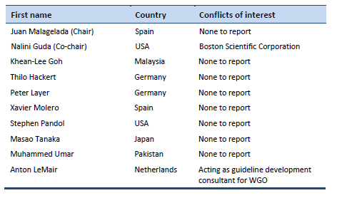
7.2  Author’s Conflict of Interest Reporting

7.3  Published Guidelines

7.3.1  International Guidelines

       2018 Gut. European evidence-based guidelines on pancreatic cystic neoplasms [29]. Available from: https://www.ncbi.nlm.nih.gov/pmc/articles/PMC5890653/

       2018 Am J Gastroenterol. ACG clinical guideline: diagnosis and management of pancreatic cysts [49].

       2017 J Am Coll Radiol. Management of incidental pancreatic cysts: a white paper of the ACR Incidental Findings Committee [50]. Available from: https://www.jacr.org/article/S1546-1440(17)30318-6/fulltext

       2017 Pancreatology. Revisions of international consensus Fukuoka guidelines for the management of IPMN of the pancreas [40].

       2015 Gastroenterology. AGA guidelines for the management of pancreatic cysts [43].

       2015 World J Gastroenterol. International guidelines for the management of pancreatic intraductal papillary mucinous neoplasms [51]. Available from: https://www.ncbi.nlm.nih.gov/pmc/articles/PMC4566378/

       2015 Ann Transl Med [Internet]. International consensus on the management of intraductal papillary mucinous neoplasm of the pancreas [45]. Available from: http://www.ncbi.nlm.nih.gov/pmc/articles/PMC4671873/

       2013 Dig Liver Dis. European experts consensus statement on cystic tumours of the pancreas [52].

       2012 Pancreatology. International consensus guidelines 2012 for the management of IPMN and MCN of the pancreas [8].

       2006 Pancreatology. International consensus guidelines for management of intraductal papillary mucinous neoplasms and mucinous cystic neoplasms of the pancreas [53].

7.3.2  Regional and Other Guidelines

       2015 J Hepatobiliary Pancreat Sci. Revised Japanese guidelines for the management of acute pancreatitis 2015: revised concepts and updated points [54].

       2015 Gastroenterology. American Gastroenterological Association technical review on the diagnosis and management of asymptomatic neoplastic pancreatic cysts [55].

       2015 Gastroenterology. AGA Institute guideline on the diagnosis and management of asymptomatic neoplastic pancreatic cysts [41].

       2015 Gastroenterology. AGA guidelines for the management of pancreatic cysts [43].

       2015 Ann Oncol. Cancer of the pancreas: ESMO clinical practice guidelines for diagnosis, treatment and follow-up [56].

       2015 Am J Gastroenterol. ACG clinical guideline: genetic testing and management of hereditary gastrointestinal cancer syndromes [57].

       2014 Saudi Med J. Saudi Oncology Society clinical management guideline series. Pancreatic cancer [58].

       2014 RöFo. S3 guideline for chronic pancreatitis—diagnosis, classification and therapy for the radiologist [59].

       2014 Pancreas. American Pancreatic Association practice guidelines in chronic pancreatitis: evidence-based report on diagnostic guidelines [60].

       2014 Jpn J Clin Oncol. EBM-based clinical guidelines for pancreatic cancer (2013) issued by the Japan Pancreas Society: a synopsis [61].

       2014 Dig Liver Dis. Italian consensus guidelines for the diagnostic work-up and follow-up of cystic pancreatic neoplasms [62].

       2014 Diagn Cytopathol. Utilization of ancillary studies in the cytologic diagnosis of biliary and pancreatic lesions: the Papanicolaou Society of Cytopathology guidelines for pancreatobiliary cytology [63].

       2014 Diagn Cytopathol. Postbrushing and fine-needle aspiration biopsy follow-up and treatment options for patients with pancreatobiliary lesions: the Papanicolaou Society of Cytopathology guidelines [64].

       2014 Diagn Cytopathol. Standardized terminology and nomenclature for pancreatobiliary cytology: the Papanicolaou Society of Cytopathology guidelines [65].

       2014 Cancer Cytopathol. Guidelines for pancreaticobiliary cytology from the Papanicolaou Society of Cytopathology: a review [66].

       2013 Am J Gastroenterol. American College of Gastroenterology guideline: management of acute pancreatitis [67].

       2012 J Natl Compr Canc Netw. Pancreatic adenocarcinoma, version 2.2012: featured updates to the NCCN Guidelines [68].

       2012 Ann Oncol. Pancreatic adenocarcinoma: ESMO-ESDO clinical practice guidelines for diagnosis, treatment and follow-up [69].

       2008 Ann Oncol. Neuroendocrine gastro-entero-pancreatic tumors: ESMO clinical practice guidelines for diagnosis, treatment and follow-up [70].

       2005 Ann Surg Oncol. Treatment guidelines for branch duct type intraductal papillary mucinous neoplasms of the pancreas: when can we operate or observe? [71].

       2004 Gastrointest Endosc. ASGE guideline: The role of endoscopy in the diagnosis and the management of cystic lesions and inflammatory fluid collections of the pancreas [72].

       Am J Surg Pathol. An illustrated consensus on the classification of pancreatic intraepithelial neoplasia and intraductal papillary mucinous neoplasms [73].

7.4 References

1.        Ohno E, Hirooka Y, Kawashima H, Ishikawa T, Kanamori A, Ishikawa H, et al. Natural history of pancreatic cystic lesions: A multicenter prospective observational study for evaluating the risk of pancreatic cancer. J Gastroenterol Hepatol. 2018 Jan;33(1):320–8.

2.        Chang YR, Park JK, Jang J-Y, Kwon W, Yoon JH, Kim S-W. Incidental pancreatic cystic neoplasms in an asymptomatic healthy population of 21,745 individuals: Large-scale, single-center cohort study. Medicine (Baltimore). 2016 Dec;95(51):e5535.

3.        Basturk O, Hong S-M, Wood LD, Adsay NV, Albores-Saavedra J, Biankin AV, et al. A revised classification system and recommendations from the Baltimore consensus meeting for neoplastic precursor lesions in the pancreas. Am J Surg Pathol. 2015 Dec;39(12):1730–41.

4.        de Pretis N, Mukewar S, Aryal-Khanal A, Bi Y, Takahashi N, Chari S. Pancreatic cysts: diagnostic accuracy and risk of inappropriate resections. Pancreatol Off J Int Assoc Pancreatol IAP Al. 2017 Apr;17(2):267–72.

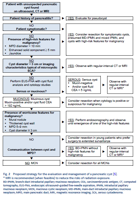
5.        Stark A, Donahue TR, Reber HA, Hines OJ. Pancreatic cyst disease: a review. JAMA. 2016 May 3;315(17):1882–93.

6.        Gaujoux S, Brennan MF, Gonen M, D’Angelica MI, DeMatteo R, Fong Y, et al. Cystic lesions of the pancreas: changes in the presentation and management of 1,424 patients at a single institution over a 15-year time period. J Am Coll Surg. 2011 Apr;212(4):590–600; discussion 600-603.

7.        Karoumpalis I, Christodoulou DK. Cystic lesions of the pancreas. Ann Gastroenterol. 2016 Jun;29(2):155–61.

8.        Tanaka M, Fernández-del Castillo C, Adsay V, Chari S, Falconi M, Jang J-Y, et al. International consensus guidelines 2012 for the management of IPMN and MCN of the pancreas. Pancreatology. 2012 Jun;12(3):183–97.

9.        Khalid A, Brugge W. ACG practice guidelines for the diagnosis and management of neoplastic pancreatic cysts. Am J Gastroenterol. 2007 Oct;102(10):2339–49.

10.      Aaltonen L, Hamilton S, Lambert R. World Health Organization classification of tumours: pathology and genetics of tumours of the digestive system. In: World Health Organization Classification of Tumours. 2000. p. 217–51.

11.      Levy MJ. Pancreatic cysts. Gastrointest Endosc. 2009 Feb;69(2 Suppl):S110-116.

12.      Lloyd R, Osamura R, Klöppel G, Rosai J. WHO classification of tumours of endocrine organs [Internet]. 4th ed. WHO IARC; 2017 [cited 2018 Apr 27]. 355 p. (IARC WHO Classification of Tumours (Book 10); vol. 10). Available from: http://publications.iarc.fr/Book-And-Report-Series/Who-Iarc-Classification-Of-Tumours/Who-Classification-Of-Tumours-Of-Endocrine-Organs-2017

13.      Guilmette JM, Nosé V. Neoplasms of the neuroendocrine pancreas: an update in the classification, definition, and molecular genetic advances. Adv Anat Pathol. 2018 Jun 14;

14.      Maire F, Couvelard A, Palazzo L, Aubert A, Vullierme M-P, Rebours V, et al. Pancreatic intraepithelial neoplasia in patients with intraductal papillary mucinous neoplasms: the interest of endoscopic ultrasonography. Pancreas. 2013 Nov;42(8):1262–6.

15.      Tsutsumi K, Ohtsuka T, Oda Y, Sadakari Y, Mori Y, Aishima S, et al. A history of acute pancreatitis in intraductal papillary mucinous neoplasms of the pancreas is a potential predictive factor for malignant papillary subtype. Pancreatology. 2010;10(6):707–12.

16.      Traverso LW, Moriya T, Hashimoto Y. Intraductal papillary mucinous neoplasms of the pancreas: making a disposition using the natural history. Curr Gastroenterol Rep. 2012 Apr;14(2):106–11.

17.      Nilsson LN, Keane MG, Shamali A, Millastre Bocos J, Marijinissen van Zanten M, Antila A, et al. Nature and management of pancreatic mucinous cystic neoplasm (MCN): A systematic review of the literature. Pancreatology. 2016 Dec;16(6):1028–36.

18.      Schmid RM, Siveke JT. Approach to cystic lesions of the pancreas. Wien Med Wochenschr 1946. 2014 Feb;164(3–4):44–50.

19.      Lévy P, Rebours V. Differential diagnosis of cystic pancreatic lesions including the usefulness of biomarkers. Viszeralmedizin. 2015 Feb;31(1):7–13.

20.      Al-Haddad MA, Kowalski T, Siddiqui A, Mertz HR, Mallat D, Haddad N, et al. Integrated molecular pathology accurately determines the malignant potential of pancreatic cysts. Endoscopy. 2015 Feb;47(2):136–42.

21.      Morris-Stiff G, Lentz G, Chalikonda S, Johnson M, Biscotti C, Stevens T, et al. Pancreatic cyst aspiration analysis for cystic neoplasms: mucin or carcinoembryonic antigen—which is better? Surgery. 2010 Oct;148(4):638–44; discussion 644-645.

22.      Rockacy M, Khalid A. Update on pancreatic cyst fluid analysis. Ann Gastroenterol. 2013;26(2):122–7.

23.      Leung KK, Ross WA, Evans D, Fleming J, Lin E, Tamm EP, et al. Pancreatic cystic neoplasm: the role of cyst morphology, cyst fluid analysis, and expectant management. Ann Surg Oncol. 2009 Oct;16(10):2818–24.

24.      Brugge WR, Lewandrowski K, Lee-Lewandrowski E, Centeno BA, Szydlo T, Regan S, et al. Diagnosis of pancreatic cystic neoplasms: a report of the cooperative pancreatic cyst study. Gastroenterology. 2004 May;126(5):1330–6.

25.      Bick BL, Enders FT, Levy MJ, Zhang L, Henry MR, Abu Dayyeh BK, et al. The string sign for diagnosis of mucinous pancreatic cysts. Endoscopy. 2015 Jul;47(7):626–31.

26.      Maker AV, Lee LS, Raut CP, Clancy TE, Swanson RS. Cytology from pancreatic cysts has marginal utility in surgical decision-making. Ann Surg Oncol. 2008 Nov;15(11):3187–92.

27.      Hong S-KS, Loren DE, Rogart JN, Siddiqui AA, Sendecki JA, Bibbo M, et al. Targeted cyst wall puncture and aspiration during EUS-FNA increases the diagnostic yield of premalignant and malignant pancreatic cysts. Gastrointest Endosc. 2012 Apr;75(4):775–82.

28.      Chiang AL, Lee LS. Clinical approach to incidental pancreatic cysts. World J Gastroenterol. 2016 Jan 21;22(3):1236–45.

29.      European Study Group on Cystic Tumours of the Pancreas. European evidence-based guidelines on pancreatic cystic neoplasms. Gut. 2018 May;67(5):789–804.

30.      Hackert T, Fritz S, Klauss M, Bergmann F, Hinz U, Strobel O, et al. Main-duct intraductal papillary mucinous nNeoplasm: high cancer risk in duct diameter of 5 to 9 mm. Ann Surg. 2015 Nov;262(5):875–80; discussion 880-881.

31.      Morales-Oyarvide V, Mino-Kenudson M, Ferrone CR, Gonzalez-Gonzalez LA, Warshaw AL, Lillemoe KD, et al. Acute pancreatitis in intraductal papillary mucinous neoplasms: A common predictor of malignant intestinal subtype. Surgery. 2015 Nov;158(5):1219–25.

32.      Konings ICAW, Harinck F, Poley J-W, Aalfs CM, van Rens A, Krak NC, et al. Prevalence and progression of pancreatic cystic precursor lesions differ between groups at high risk of developing pancreatic cancer. Pancreas. 2017 Jan;46(1):28–34.

33.      Capurso G, Boccia S, Salvia R, Del Chiaro M, Frulloni L, Arcidiacono PG, et al. Risk factors for intraductal papillary mucinous neoplasm (IPMN) of the pancreas: a multicentre case-control study. Am J Gastroenterol. 2013 Jun;108(6):1003–9.

34.      Ohtsuka T, Kono H, Nagayoshi Y, Mori Y, Tsutsumi K, Sadakari Y, et al. An increase in the number of predictive factors augments the likelihood of malignancy in branch duct intraductal papillary mucinous neoplasm of the pancreas. Surgery. 2012 Jan;151(1):76–83.

35.      Perez-Johnston R, Narin O, Mino-Kenudson M, Ingkakul T, Warshaw AL, Fernandez-Del Castillo C, et al. Frequency and significance of calcification in IPMN. Pancreatology. 2013 Feb;13(1):43–7.

36.      Yagi Y, Masuda A, Zen Y, Takenaka M, Toyama H, Sofue K, et al. Predictive value of low serum pancreatic enzymes in invasive intraductal papillary mucinous neoplasms. Pancreatol Off J Int Assoc Pancreatol IAP Al. 2016 Oct;16(5):893–9.

37.      Fritz S, Hackert T, Hinz U, Hartwig W, Büchler MW, Werner J. Role of serum carbohydrate antigen 19-9 and carcinoembryonic antigen in distinguishing between benign and invasive intraductal papillary mucinous neoplasm of the pancreas. Br J Surg. 2011 Jan;98(1):104–10.

38.      Chang Y-T, Tien Y-W, Jeng Y-M, Yang C-Y, Liang P-C, Wong J-M, et al. Overweight increases the risk of malignancy in patients with pancreatic mucinous cystic neoplasms. Medicine (Baltimore). 2015 May;94(20):e797.

39.      Lafemina J, Katabi N, Klimstra D, Correa-Gallego C, Gaujoux S, Kingham TP, et al. Malignant progression in IPMN: a cohort analysis of patients initially selected for resection or observation. Ann Surg Oncol. 2013 Feb;20(2):440–7.

40.      Tanaka M, Fernández-del Castillo C, Kamisawa T, Jang JY, Levy P, Ohtsuka T, et al. Revisions of international consensus Fukuoka guidelines for the management of IPMN of the pancreas. Pancreatology. 2017 Sep;17(5):738–53.

41.      Vege SS, Ziring B, Jain R, Moayyedi P, Clinical Guidelines Committee, American Gastroenterology Association. American Gastroenterological Association institute guideline on the diagnosis and management of asymptomatic neoplastic pancreatic cysts. Gastroenterology. 2015 Apr;148(4):819–22; quize12-13.

42.      Sahani DV, Kambadakone A, Macari M, Takahashi N, Chari S, Fernandez-del Castillo C. Diagnosis and management of cystic pancreatic lesions. AJR Am J Roentgenol. 2013 Feb;200(2):343–54.

43.      Lennon AM, Ahuja N, Wolfgang CL. AGA guidelines for the management of pancreatic cysts. Gastroenterology. 2015 Sep;149(3):825.

44.      Heckler M, Michalski CW, Schaefle S, Kaiser J, Büchler MW, Hackert T. The Sendai and Fukuoka consensus criteria for the management of branch duct IPMN—a meta-analysis on their accuracy. Pancreatology. 2017 Mar;17(2):255–62.

45.      Tanaka M. International consensus on the management of intraductal papillary mucinous neoplasm of the pancreas. Ann Transl Med. 2015 Nov;3(19):286.

46.      Choi SH, Park SH, Kim KW, Lee JY, Lee SS. Progression of unresected intraductal papillary mucinous neoplasms of the pancreas to cancer: a systematic review and meta-analysis. Clin Gastroenterol Hepatol. 2017 Oct;15(10):1509-1520.e4.

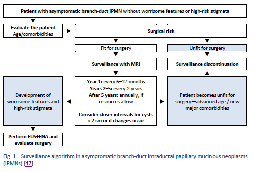
47.      Crippa S, Pezzilli R, Bissolati M, Capurso G, Romano L, Brunori MP, et al. Active surveillance beyond 5 years is required for presumed branch-duct intraductal papillary mucinous neoplasms undergoing non-operative management. Am J Gastroenterol. 2017 Jul;112(7):1153–61.

48.      Anand N, Sampath K, Wu BU. Cyst features and risk of malignancy in intraductal papillary mucinous neoplasms of the pancreas: a meta-analysis. Clin Gastroenterol Hepatol. 2013 Aug;11(8):913–21; quiz e59-60.

49.      Elta GH, Enestvedt BK, Sauer BG, Lennon AM. ACG clinical guideline: diagnosis and management of pancreatic cysts. Am J Gastroenterol. 2018;113(4):464–79.

50.      Megibow AJ, Baker ME, Morgan DE, Kamel IR, Sahani DV, Newman E, et al. Management of incidental pancreatic cysts: a white paper of the ACR Incidental Findings Committee. J Am Coll Radiol JACR. 2017 Jul;14(7):911–23.

51.      Goh BK. International guidelines for the management of pancreatic intraductal papillary mucinous neoplasms. World J Gastroenterol WJG. 2015 Sep 14;21(34):9833–7.

52.      Del Chiaro M, Verbeke C, Salvia R, Klöppel G, Werner J, McKay C, et al. European experts consensus statement on cystic tumours of the pancreas. Dig Liver Dis. 2013 Sep;45(9):703–11.

53.      Tanaka M, Chari S, Adsay V, Fernandez-del Castillo C, Falconi M, Shimizu M, et al. International consensus guidelines for management of intraductal papillary mucinous neoplasms and mucinous cystic neoplasms of the pancreas. Pancreatology. 2006;6(1–2):17–32.

54.      Isaji S, Takada T, Mayumi T, Yoshida M, Wada K, Yokoe M, et al. Revised Japanese guidelines for the management of acute pancreatitis 2015: revised concepts and updated points. J Hepato-Biliary-Pancreat Sci. 2015 Jun;22(6):433–45.

55.      Scheiman JM, Hwang JH, Moayyedi P. American Gastroenterological Association technical review on the diagnosis and management of asymptomatic neoplastic pancreatic cysts. Gastroenterology. 2015 Apr;148(4):824-848.e22.

56.      Ducreux M, Cuhna AS, Caramella C, Hollebecque A, Burtin P, Goéré D, et al. Cancer of the pancreas: ESMO Clinical Practice Guidelines for diagnosis, treatment and follow-up. Ann Oncol Off J Eur Soc Med Oncol. 2015 Sep;26 Suppl 5:v56-68.

57.      Syngal S, Brand RE, Church JM, Giardiello FM, Hampel HL, Burt RW, et al. ACG clinical guideline: genetic testing and management of hereditary gastrointestinal cancer syndromes. Am J Gastroenterol. 2015 Feb;110(2):223–62; quiz 263.

58.      Rahal MM, Bazarbashi SN, Kandil MS, Al-Shehri AS, Alzahrani AM, Aljubran AH, et al. Saudi Oncology Society clinical management guideline series. Pancreatic cancer 2014. Saudi Med J. 2014 Dec;35(12):1534–7.

59.      Schreyer AG, Jung M, Riemann JF, Niessen C, Pregler B, Grenacher L, et al. S3 guideline for chronic pancreatitis—diagnosis, classification and therapy for the radiologist. RöFo. 2014 Nov;186(11):1002–8.

60.      Conwell DL, Lee LS, Yadav D, Longnecker DS, Miller FH, Mortele KJ, et al. American Pancreatic Association practice guidelines in chronic pancreatitis: evidence-based report on diagnostic guidelines. Pancreas. 2014 Nov;43(8):1143–62.

61.      Yamaguchi K, Okusaka T, Shimizu K, Furuse J, Ito Y, Hanada K, et al. EBM-based clinical guidelines for pancreatic cancer (2013) issued by the Japan Pancreas Society: a synopsis. Jpn J Clin Oncol. 2014 Oct;44(10):883–8.

62.      Italian Association of Hospital Gastroenterologists and Endoscopists, Italian Association for the Study of the Pancreas, Buscarini E, Pezzilli R, Cannizzaro R, De Angelis C, et al. Italian consensus guidelines for the diagnostic work-up and follow-up of cystic pancreatic neoplasms. Dig Liver Dis. 2014 Jun;46(6):479–93.

63.      Layfield LJ, Ehya H, Filie AC, Hruban RH, Jhala N, Joseph L, et al. Utilization of ancillary studies in the cytologic diagnosis of biliary and pancreatic lesions: the Papanicolaou Society of Cytopathology guidelines for pancreatobiliary cytology. Diagn Cytopathol. 2014 Apr;42(4):351–62.

64.      Kurtycz D, Tabatabai ZL, Michaels C, Young N, Schmidt CM, Farrell J, et al. Postbrushing and fine-needle aspiration biopsy follow-up and treatment options for patients with pancreatobiliary lesions: the Papanicolaou Society of Cytopathology guidelines. Diagn Cytopathol. 2014 Apr;42(4):363–71.

65.      Pitman MB, Centeno BA, Ali SZ, Genevay M, Stelow E, Mino-Kenudson M, et al. Standardized terminology and nomenclature for pancreatobiliary cytology: the Papanicolaou Society of Cytopathology guidelines. Diagn Cytopathol. 2014 Apr;42(4):338–50.

66.      Pitman MB, Layfield LJ. Guidelines for pancreaticobiliary cytology from the Papanicolaou Society of Cytopathology: A review. Cancer Cytopathol. 2014 Jun;122(6):399–411.

67.      Tenner S, Baillie J, DeWitt J, Vege SS, American College of Gastroenterology. American College of Gastroenterology guideline: management of acute pancreatitis. Am J Gastroenterol. 2013 Sep;108(9):1400–15; 1416.

68.      Tempero MA, Arnoletti JP, Behrman SW, Ben-Josef E, Benson AB, Casper ES, et al. Pancreatic adenocarcinoma, version 2.2012: featured updates to the NCCN Guidelines. J Natl Compr Cancer Netw JNCCN. 2012 Jun 1;10(6):703–13.

69.      Seufferlein T, Bachet JB, Van Cutsem E, Rougier P, ESMO Guidelines Working Group. Pancreatic adenocarcinoma: ESMO-ESDO clinical practice guidelines for diagnosis, treatment and follow-up. Ann Oncol. 2012 Oct;23 Suppl 7:vii33-40.

70.      Öberg K, Knigge U, Kwekkeboom D, Perren A, ESMO Guidelines Working Group. Neuroendocrine gastro-entero-pancreatic tumors: ESMO Clinical Practice Guidelines for diagnosis, treatment and follow-up. Ann Oncol. 2012 Oct;23 Suppl 7:vii124-130.

71.      Jang J-Y, Kim S-W, Lee SE, Yang SH, Lee KU, Lee YJ, et al. Treatment guidelines for branch duct type intraductal papillary mucinous neoplasms of the pancreas: when can we operate or observe? Ann Surg Oncol. 2008 Jan;15(1):199–205.

72.      Jacobson BC, Baron TH, Adler DG, Davila RE, Egan J, Hirota WK, et al. ASGE guideline: the role of endoscopy in the diagnosis and the management of cystic lesions and inflammatory fluid collections of the pancreas. Gastrointest Endosc. 2005 Mar;61(3):363–70.

73.      Hruban RH, Takaori K, Klimstra DS, Adsay NV, Albores-Saavedra J, Biankin AV, et al. An illustrated consensus on the classification of pancreatic intraepithelial neoplasia and intraductal papillary mucinous neoplasms. Am J Surg Pathol. 2004 Aug;28(8):977–87.


Share

Website Disclaimer
X
Global Guidelines Survey

Cookie Notice

We use cookies to ensure you the best experience on our website. Your acceptance helps ensure that experience happens. To learn more, please visit our Privacy Notice.

OK